Radiologic technology programs offer a dynamic pathway into a rewarding healthcare career. These programs provide comprehensive training in the principles and practices of medical imaging, equipping graduates with the skills and knowledge to excel in a rapidly evolving field. Students delve into a diverse curriculum encompassing theoretical coursework and extensive hands-on clinical rotations, preparing them for the demands of professional practice. The rigorous training ensures competency in various imaging modalities, emphasizing both technical proficiency and patient care.
A successful completion of a radiologic technology program leads to certification and licensure, opening doors to a wide array of career opportunities within hospitals, clinics, and specialized imaging centers. The program’s emphasis on ethical considerations, radiation safety, and patient interaction ensures graduates are well-prepared not only technically but also professionally, committed to providing high-quality care in a responsible and compassionate manner.
Program Overview
Becoming a qualified radiologic technologist requires rigorous training and a commitment to mastering both theoretical knowledge and practical skills. This program provides a comprehensive education in the field, preparing graduates for entry-level positions in a variety of healthcare settings. The curriculum blends classroom learning with extensive hands-on experience in clinical settings.
The curriculum of a typical radiologic technology program is structured to provide a strong foundation in the principles of radiation physics, patient care, and imaging techniques. Students learn to operate sophisticated medical imaging equipment, interpret images, and apply radiation safety protocols. The program emphasizes the importance of ethical considerations and professional conduct within the medical field.
Required Coursework
The coursework component of the program covers a wide range of subjects essential for competent practice. These include anatomy and physiology, radiation physics, radiographic imaging techniques, radiation protection and safety, medical ethics, patient care, and image evaluation. Specific courses might also cover advanced imaging modalities such as computed tomography (CT), magnetic resonance imaging (MRI), and fluoroscopy. Students will engage in extensive didactic learning, laboratory sessions, and simulated clinical experiences to solidify their understanding of these core concepts. Successful completion of all coursework is a prerequisite for clinical rotations.
Clinical Rotations
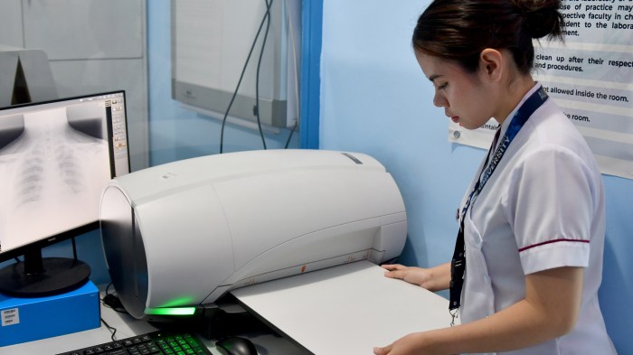
Clinical rotations are an integral part of the program, providing students with invaluable practical experience under the supervision of experienced radiologic technologists. These rotations typically take place in various healthcare settings, including hospitals, clinics, and outpatient imaging centers. Students gain hands-on experience in performing a variety of imaging procedures, interacting with patients, and working as part of a healthcare team. The clinical rotations allow students to apply the theoretical knowledge acquired in the classroom to real-world scenarios, fostering critical thinking and problem-solving skills. A comprehensive evaluation of performance during these rotations is crucial for program completion. Examples of clinical rotations might include general radiography, fluoroscopy, CT scanning, and MRI scanning, depending on the specific program and available facilities.
Accreditation Process
Accreditation ensures that radiologic technology programs meet established standards of quality and excellence. The Joint Review Committee on Education in Radiologic Technology (JRCERT) is the primary accrediting body in the United States. The accreditation process involves a rigorous review of the program’s curriculum, faculty qualifications, facilities, and clinical affiliations. Programs must meet specific criteria related to educational resources, student learning outcomes, and clinical training opportunities. Maintaining accreditation requires ongoing compliance with JRCERT standards and periodic self-studies and site visits by JRCERT representatives. Graduates from accredited programs are more likely to be eligible for certification and licensure, enhancing their career prospects. For example, a program undergoing accreditation might submit a comprehensive self-study report detailing all aspects of its operations, followed by an on-site visit by JRCERT evaluators to verify the information provided and assess the program’s overall quality.
Admission Requirements
Gaining admission to a Radiologic Technology program requires meeting specific academic and sometimes personal qualifications. These requirements vary between institutions, but generally involve a combination of academic performance, prerequisite coursework, and sometimes additional assessments. Understanding these requirements is crucial for prospective students to successfully apply and begin their studies.
Admission requirements for radiologic technology programs are designed to ensure applicants possess the necessary foundational knowledge and skills to succeed in the demanding curriculum. Programs aim to select candidates with a demonstrated aptitude for science, a commitment to learning, and the potential to become competent and compassionate healthcare professionals. The competitive nature of many programs often leads to higher admission standards, making thorough preparation essential.
Typical Admission Requirements
A common set of admission requirements typically includes a high school diploma or GED, a minimum GPA (often a 2.5 or higher), and successful completion of specific prerequisite courses. Many programs also require submission of official transcripts, letters of recommendation, and a completed application form. Some institutions may conduct interviews as part of the selection process. Additionally, criminal background checks and drug screenings are frequently required due to the sensitive nature of patient care and access to controlled substances.
Comparison of Admission Requirements Across Programs
While the core requirements mentioned above are prevalent, variations exist between different radiologic technology programs. For example, some programs may require a higher minimum GPA, specific science courses beyond the general prerequisites (such as anatomy and physiology), or even standardized test scores like the SAT or ACT. The rigor of prerequisite courses can also differ, with some programs demanding higher-level courses or a greater number of credit hours. Furthermore, the application process itself may vary, with some programs utilizing online application portals while others require paper applications. One program might prioritize clinical experience, while another might emphasize academic achievement. It’s crucial to carefully review the specific admission requirements of each program a prospective student is interested in.
Importance of Prerequisite Courses
Prerequisite courses, such as anatomy, physiology, and medical terminology, are essential for success in a radiologic technology program. These courses provide the foundational knowledge necessary to understand the human body, its systems, and the medical language used in healthcare settings. A strong grasp of these subjects enables students to comprehend complex radiological concepts, interpret medical images effectively, and communicate clearly with healthcare professionals. Failing to demonstrate proficiency in these areas may result in difficulty understanding the program’s advanced material, potentially impacting a student’s ability to complete the program successfully. Many programs may even have minimum grade requirements for these prerequisites, often a “C” or better, further highlighting their importance.
Career Opportunities
A career in radiologic technology offers a diverse range of opportunities for graduates, providing excellent job security and a rewarding path in the healthcare field. The demand for skilled radiologic technologists is consistently high, driven by an aging population and advancements in medical imaging technology. This section explores the various career paths, salary expectations, and specialized areas within the field.
Graduates of radiologic technology programs are highly sought after by hospitals, clinics, outpatient imaging centers, and physician’s offices. The job outlook is positive, with projections indicating continued growth in the coming years. Salaries are competitive and vary depending on experience, location, specialization, and employer.
Job Outlook and Salary Expectations
The Bureau of Labor Statistics projects substantial growth in the healthcare industry, leading to a strong demand for radiologic technologists. While precise salary figures fluctuate based on geographic location and experience, entry-level positions typically offer a competitive starting salary, with significant potential for advancement and increased earnings as technologists gain experience and specialize. For instance, a recent survey indicated that entry-level radiologic technologists in major metropolitan areas might earn between $50,000 and $65,000 annually, while experienced specialists can command significantly higher salaries. These figures can vary considerably based on location, employer size, and individual skills.
Specialized Areas in Radiologic Technology
Radiologic technology offers various specialized areas, allowing technologists to focus their skills and expertise in specific imaging modalities or patient populations. This specialization often leads to higher earning potential and greater job satisfaction.
| Specialization | Job Duties | Required Skills | Salary Range (USD) |
|---|---|---|---|
| Computed Tomography (CT) | Operating CT scanners, preparing patients, performing scans, and reconstructing images. | Strong anatomical knowledge, proficiency in CT scanner operation, excellent patient communication skills. | $60,000 – $85,000 |
| Magnetic Resonance Imaging (MRI) | Operating MRI scanners, preparing patients, performing scans, and analyzing images. | Detailed understanding of physics and MRI principles, excellent patient communication skills, ability to work independently. | $65,000 – $90,000 |
| Mammography | Performing mammograms, interpreting images (with additional certification), and providing patient education. | Exceptional attention to detail, understanding of breast anatomy and pathology, compassionate patient care skills. | $55,000 – $75,000 |
| Interventional Radiology | Assisting physicians during minimally invasive procedures, operating imaging equipment, and managing sterile fields. | Strong understanding of anatomy and physiology, excellent sterile technique, ability to work effectively under pressure. | $70,000 – $100,000+ |
Educational Resources
Success in a radiologic technology program hinges on access to a variety of high-quality educational resources. These resources extend beyond lectures and textbooks, encompassing a rich blend of learning tools designed to foster both theoretical understanding and practical skills. A well-rounded program provides students with the support they need to thrive in this demanding field.
A robust radiologic technology program offers a diverse range of learning materials and support systems. These resources are carefully selected to complement different learning styles and ensure students develop the necessary competencies.
Types of Educational Resources
Radiologic technology programs utilize a multifaceted approach to education, employing various resources to enhance student learning. These resources are designed to cater to diverse learning styles and ensure a comprehensive understanding of the subject matter. Textbooks provide foundational knowledge, while online learning platforms offer flexibility and supplementary materials. Access to state-of-the-art equipment and experienced instructors facilitates hands-on training and practical application of learned concepts. Furthermore, many programs incorporate simulation labs, allowing students to practice procedures in a safe and controlled environment before working with real patients. Finally, regular assessments, including quizzes, exams, and clinical evaluations, help students track their progress and identify areas for improvement.
Online versus On-Campus Learning
Online and on-campus learning models each present distinct advantages and disadvantages within the context of radiologic technology education. On-campus programs offer the benefit of direct interaction with instructors and peers, fostering a collaborative learning environment and immediate access to equipment and facilities. However, on-campus learning often necessitates a rigid schedule and geographical proximity to the institution. In contrast, online programs provide greater flexibility and accessibility, allowing students to learn at their own pace and from any location with an internet connection. However, online learning can lack the hands-on experience and immediate feedback provided by an on-campus setting. The optimal learning model depends on individual learning styles, personal circumstances, and program structure. Many programs now offer hybrid models that combine the benefits of both approaches.
Simulation and Hands-on Training
Simulation and hands-on training are integral components of effective radiologic technology education. Simulation labs provide a safe and controlled environment for students to practice various procedures, including positioning patients, operating equipment, and interpreting images, before transitioning to clinical settings. This minimizes risks associated with real-patient interactions and allows students to refine their skills through repeated practice and instructor feedback. Hands-on training in clinical settings, under the supervision of experienced professionals, is equally crucial. This practical experience allows students to apply their theoretical knowledge in real-world scenarios, develop critical thinking skills, and gain confidence in their abilities. The combination of simulation and hands-on training provides a well-rounded learning experience, preparing students for the demands of a professional career in radiologic technology.
Technological Advancements
The field of radiologic technology is experiencing rapid evolution, driven by continuous advancements in imaging technology. These improvements not only enhance the quality and speed of diagnostic imaging but also significantly impact patient care and the overall efficiency of healthcare systems. This constant innovation necessitates ongoing adaptation and integration within radiologic technology programs to ensure graduates are equipped with the skills and knowledge required for contemporary practice.
New technologies are integrated into radiologic technology programs through a variety of methods. Curricula are regularly updated to reflect the latest advancements, often incorporating hands-on training with cutting-edge equipment. Lectures, simulations, and clinical rotations provide students with practical experience using these new technologies, preparing them for the challenges and opportunities of the modern imaging environment. Furthermore, many programs incorporate continuing education opportunities and professional development workshops to ensure that graduates remain current with the latest technological trends throughout their careers.
Examples of Cutting-Edge Technologies in Radiologic Imaging
Modern radiologic technology utilizes a range of sophisticated imaging modalities. These technologies offer improved image quality, reduced radiation exposure, and enhanced diagnostic capabilities, ultimately leading to more accurate diagnoses and improved patient outcomes.
- Digital Radiography (DR): DR systems have largely replaced traditional film-based radiography. They utilize digital detectors to capture X-ray images, which are then processed and displayed on a computer monitor. This allows for immediate image review, manipulation, and electronic storage, eliminating the need for film processing and storage. DR systems often incorporate features like image stitching and advanced image processing algorithms to improve image quality and diagnostic accuracy.
- Computed Tomography (CT): CT scanners use X-rays to create detailed cross-sectional images of the body. Modern CT scanners employ multislice technology, allowing for faster scan times and improved spatial resolution. Advanced reconstruction algorithms and iterative reconstruction techniques further enhance image quality and reduce radiation dose. Examples of advancements include dual-energy CT, which allows for the differentiation of materials with similar densities, and spectral CT, offering improved tissue characterization.
- Magnetic Resonance Imaging (MRI): MRI uses powerful magnets and radio waves to generate detailed images of internal organs and tissues. Advances in MRI technology include higher field strength magnets, which provide improved image resolution and contrast. Functional MRI (fMRI) techniques allow for the study of brain activity, while diffusion tensor imaging (DTI) provides information about the white matter tracts in the brain. These advancements have expanded the clinical applications of MRI, allowing for more precise diagnoses and treatment planning.
- Nuclear Medicine Imaging: Nuclear medicine techniques utilize radioactive tracers to visualize physiological processes within the body. Positron emission tomography (PET) and single-photon emission computed tomography (SPECT) are two widely used nuclear medicine imaging modalities. Advances in PET/CT and SPECT/CT fusion imaging combine the functional information from nuclear medicine with the anatomical detail from CT, providing comprehensive diagnostic information. The development of new radiotracers with improved specificity and sensitivity continues to expand the capabilities of nuclear medicine imaging.
- Ultrasound: Ultrasound uses high-frequency sound waves to create images of internal organs and tissues. Advances in ultrasound technology include higher-frequency transducers, which provide improved image resolution, and advanced imaging techniques such as elastography, which assesses the stiffness of tissues. Three-dimensional and four-dimensional ultrasound provide more detailed anatomical information and can be used to visualize fetal development.
Professional Organizations
Joining a professional organization is a vital step for radiologic technologists seeking to advance their careers and contribute to the field’s growth. These organizations offer a wealth of resources, networking opportunities, and advocacy efforts that benefit both individual members and the profession as a whole. They play a crucial role in setting standards, promoting continuing education, and shaping the future of radiologic technology.
Professional organizations provide a platform for radiologic technologists to connect with colleagues, share experiences, and learn from experts. Membership often includes access to journals, conferences, and online resources, fostering professional development and staying current with the latest advancements in the field. These organizations also actively advocate for the interests of radiologic technologists, influencing policy decisions and ensuring fair compensation and working conditions.
Key Professional Organizations for Radiologic Technologists
The American Society of Radiologic Technologists (ASRT) is the largest professional organization for radiologic technologists in the United States. It offers a wide range of benefits, including continuing education opportunities, professional liability insurance, and advocacy for its members. Another significant organization is the American Registry of Radiologic Technologists (ARRT), which is responsible for credentialing and certifying radiologic technologists. Membership in organizations like the Association of Educators in Radiologic Technology (AEIRT) is particularly beneficial for educators and those involved in training the next generation of radiologic technologists. These organizations often collaborate on initiatives to improve the quality of education and training in the field.
Benefits of Membership in Professional Organizations
Membership in professional organizations offers numerous advantages. These include access to continuing education courses, workshops, and webinars, keeping members abreast of the latest techniques, technologies, and best practices. Networking opportunities at conferences and meetings provide chances to connect with peers, mentors, and potential employers. Many organizations also offer professional liability insurance, protecting members from potential legal issues. Furthermore, active participation in professional organizations demonstrates a commitment to professional development and enhances career prospects. Advocacy efforts undertaken by these organizations work to improve working conditions, compensation, and the overall standing of radiologic technologists within the healthcare industry. For example, ASRT actively lobbies for legislation affecting the profession, ensuring that radiologic technologists’ voices are heard in policy-making.
The Role of Professional Certifications in the Field
Professional certifications, such as those offered by the ARRT, are essential for radiologic technologists. Certification demonstrates competence and adherence to professional standards, enhancing credibility and employability. Different certifications are available, specializing in various areas of radiologic technology, such as mammography, computed tomography (CT), and magnetic resonance imaging (MRI). Obtaining and maintaining these certifications requires ongoing professional development and adherence to continuing education requirements, demonstrating a commitment to lifelong learning and maintaining high standards of practice. These certifications often translate to higher earning potential and increased career opportunities. For instance, a radiologic technologist certified in MRI may command a higher salary than one without specialized certification. Furthermore, some employers may require specific certifications for certain positions, highlighting the importance of professional credentials in securing and advancing in the field.
Ethical Considerations: Radiologic Technology Program
Radiologic technology demands a high level of ethical conduct, encompassing not only technical proficiency but also a deep commitment to patient well-being and professional integrity. The ethical responsibilities of a radiologic technologist extend beyond the technical aspects of the job, impacting patient care, professional interactions, and the overall reputation of the profession. These responsibilities are guided by professional codes of ethics and legal frameworks designed to protect patients and uphold the standards of the profession.
The cornerstone of ethical practice in radiologic technology is the unwavering commitment to patient safety and well-being. This necessitates a thorough understanding and adherence to established safety protocols, including radiation protection measures and the appropriate use of imaging techniques. Equally crucial is the ability to communicate effectively with patients, addressing their concerns and ensuring their comfort and understanding throughout the imaging process. This also includes the careful consideration of a patient’s physical and emotional state, ensuring procedures are tailored to their individual needs and capabilities.
Patient Privacy and Confidentiality
Maintaining patient privacy and confidentiality is paramount in radiologic technology. Patient information, including medical history, imaging results, and personal details, is considered protected health information (PHI) and is subject to stringent regulations under laws like HIPAA (Health Insurance Portability and Accountability Act) in the United States. Breaches of confidentiality can have severe legal and ethical consequences, damaging both the patient’s trust and the reputation of the healthcare provider. Radiologic technologists must strictly adhere to established protocols for handling and storing patient data, ensuring that access is limited to authorized personnel only. This includes secure storage of physical files, the use of encrypted electronic systems, and the responsible use of patient information within the healthcare setting. Examples of breaches could include discussing a patient’s condition with unauthorized individuals, leaving patient files unattended, or improperly disposing of patient records. Strict adherence to established protocols is essential to avoid such breaches.
Radiation Safety and Professional Ethics
Professional ethics plays a crucial role in ensuring radiation safety. Radiologic technologists are responsible for minimizing radiation exposure to both patients and themselves, adhering to the ALARA principle (“As Low As Reasonably Achievable”). This involves careful selection of imaging techniques, optimization of exposure parameters, and the use of appropriate shielding measures. Regular calibration and maintenance of equipment are essential to ensure optimal performance and minimize radiation leakage. Furthermore, continuing education in radiation safety is vital to staying abreast of advancements and best practices. Failure to adhere to radiation safety protocols not only poses a risk to patients and colleagues but also constitutes a serious ethical breach. For example, neglecting to use appropriate shielding during a procedure or failing to properly adjust the equipment settings could result in unnecessary radiation exposure to a patient, leading to potential long-term health risks. The ethical responsibility of a technologist extends to actively participating in quality assurance programs, reporting any equipment malfunctions promptly, and advocating for a safe radiation environment.
Clinical Skills
Becoming a proficient radiologic technologist requires a diverse skill set extending beyond theoretical knowledge. Practical, hands-on experience is crucial for developing the dexterity, precision, and patient interaction skills necessary for safe and effective imaging procedures. These skills are honed through rigorous clinical rotations, supervised practice, and continuous learning.
Clinical rotations provide the essential bridge between classroom learning and real-world application. Students transition from a controlled learning environment to a dynamic clinical setting, where they work alongside experienced radiologic technologists and other healthcare professionals. This immersive experience allows for the development of critical thinking, problem-solving, and teamwork skills in a practical context. Direct patient interaction, under the guidance of preceptors, allows for the refinement of communication and patient care techniques.
Essential Clinical Skills for Radiologic Technologists
The acquisition of essential clinical skills is a progressive process, building upon foundational knowledge and evolving through practical experience. These skills encompass patient assessment, positioning, radiation safety protocols, image acquisition, image evaluation, quality control, and equipment operation. Furthermore, effective communication, teamwork, and adherence to ethical standards are paramount.
Imaging Modalities and Associated Techniques
A comprehensive understanding of various imaging modalities and their associated techniques is fundamental to the role of a radiologic technologist. Each modality requires specific positioning, technical parameters, and safety protocols to ensure optimal image quality and patient safety.
- X-ray: Includes various techniques such as chest x-rays, abdominal x-rays, extremity x-rays, and fluoroscopy. Positioning is crucial to minimize superimposition and maximize visualization of the area of interest. Knowledge of radiation protection principles is essential to minimize patient exposure.
- Computed Tomography (CT): Involves scanning the body in thin slices to create detailed cross-sectional images. Precise patient positioning and the selection of appropriate scan parameters (kVp, mA, slice thickness) are vital for optimal image quality. Contrast administration techniques may be used to enhance visualization of specific structures.
- Magnetic Resonance Imaging (MRI): Uses magnetic fields and radio waves to produce high-resolution images of internal organs and tissues. Patient preparation, coil selection, and sequence parameter adjustments are crucial for producing high-quality images. Safety considerations related to magnetic fields and implanted devices must be carefully addressed.
- Mammography: Specializes in imaging the breast. Proper patient positioning and compression techniques are crucial for minimizing discomfort and maximizing image quality. Knowledge of breast anatomy and pathology is important for image interpretation.
- Fluoroscopy: Provides real-time imaging of internal structures, often used during surgical procedures or interventional radiology. Requires precise positioning and manipulation of the image intensifier to visualize the area of interest while minimizing radiation exposure.
- Ultrasound: Uses high-frequency sound waves to produce images of internal structures. Requires knowledge of anatomy, transducer selection, and image optimization techniques. Different transducer types are used depending on the area being imaged.
Patient Interaction
Effective communication and a compassionate approach are paramount in radiologic technology. Patients undergoing procedures often experience anxiety, and a skilled technologist can significantly impact their comfort and cooperation through skillful interaction. Building rapport and trust is crucial for a successful examination, leading to higher quality images and a more positive patient experience.
Successful patient interaction hinges on several key communication strategies. Empathy and sensitivity are not merely desirable traits but essential components of providing high-quality care. Understanding and acknowledging a patient’s feelings and concerns creates a safe and supportive environment, encouraging open communication and cooperation.
Effective Communication Strategies
Effective communication involves more than simply providing instructions. It requires active listening, clear and concise explanations, and sensitivity to individual needs. For example, using plain language, avoiding medical jargon, and checking for understanding are vital steps. Visual aids, such as diagrams or models, can also be helpful in explaining complex procedures. Furthermore, maintaining eye contact, using a calm and reassuring tone, and respecting personal space all contribute to a positive interaction. Involving the patient in the process, such as explaining the steps involved and answering their questions patiently, fosters trust and reduces anxiety.
Empathy and Sensitivity in Patient Care
Empathy involves understanding and sharing the feelings of another person. In radiology, this means recognizing that patients may be experiencing fear, pain, or discomfort. Sensitivity involves demonstrating awareness and respect for these feelings. For example, acknowledging a patient’s apprehension about a procedure, offering comfort measures like a blanket or pillow, and allowing time for questions demonstrate empathy and sensitivity. Addressing individual needs, such as adjusting the room temperature or providing privacy, further enhances the patient experience. Recognizing and respecting diverse cultural backgrounds and communication styles also contributes to a more inclusive and empathetic approach.
Managing Anxious or Fearful Patients
Many patients experience anxiety or fear before and during radiologic procedures. Managing these emotions requires a calm and reassuring approach. Techniques include providing clear and concise explanations of the procedure, answering questions patiently, and offering reassurance. Distraction techniques, such as engaging the patient in conversation or offering them music to listen to, can also be helpful. In cases of severe anxiety, collaboration with the referring physician or a healthcare professional specializing in anxiety management may be necessary. Furthermore, providing positive reinforcement and acknowledging the patient’s bravery throughout the procedure can significantly alleviate anxiety. For instance, a simple phrase like, “You’re doing great,” can make a difference.
Radiation Safety
Radiation safety is paramount in radiologic technology, demanding a comprehensive understanding and strict adherence to established protocols. The potential for ionizing radiation to cause harm to both patients and healthcare workers necessitates a proactive approach to minimizing exposure and ensuring the safety of all involved. This involves a multi-faceted strategy encompassing equipment, technique, and personal protective measures.
Minimizing radiation exposure relies on a combination of techniques and protective measures. The primary goal is to achieve diagnostic image quality using the lowest possible radiation dose. This “ALARA” principle – As Low As Reasonably Achievable – guides all practices. This involves optimizing technical factors such as kilovoltage peak (kVp), milliampere-seconds (mAs), and source-to-image distance (SID) to reduce unnecessary radiation while maintaining image clarity. Furthermore, the use of shielding devices like lead aprons, thyroid collars, and protective glasses for healthcare workers is crucial. For patients, appropriate shielding techniques, such as lead shields placed strategically over reproductive organs or other sensitive areas, are employed. Regular equipment calibration and quality control procedures also play a significant role in ensuring optimal performance and minimizing radiation output.
Radiation Safety Protocols
Implementing effective radiation safety protocols requires a structured approach. These protocols aim to protect both patients and personnel from unnecessary radiation exposure, ensuring compliance with all relevant regulations and best practices. Strict adherence to these guidelines is crucial for maintaining a safe working environment and minimizing the risk of long-term health consequences. The protocols encompass all aspects of the imaging process, from initial patient assessment to image acquisition and post-procedure analysis. Comprehensive training and regular refresher courses are essential to ensure that all staff are knowledgeable about and proficient in these protocols.
Methods for Minimizing Radiation Exposure
Several methods are used to minimize radiation exposure. These methods can be categorized into three main areas: optimization of technical factors, use of protective barriers, and implementation of effective radiation safety practices. Optimizing technical factors involves adjusting the kVp, mAs, and SID to achieve the best image quality with the lowest possible radiation dose. The use of protective barriers includes lead aprons, thyroid shields, and other shielding devices to reduce radiation exposure to personnel and patients. Finally, effective radiation safety practices encompass proper patient positioning, collimation, and the use of image receptors with high detective quantum efficiency (DQE). Regular monitoring of radiation levels and adherence to ALARA principles are also crucial.
Flowchart for Ensuring Radiation Safety During a Procedure
The following flowchart illustrates the steps involved in ensuring radiation safety during a radiographic procedure. Each step is crucial in minimizing radiation exposure and maintaining a safe environment for both patients and healthcare workers. Deviation from these steps could result in increased radiation exposure and potential harm.
[Diagram would be inserted here. Description: A flowchart showing the following steps: 1. Pre-procedure planning (including patient history, assessment of radiation risk factors, and selection of appropriate imaging technique); 2. Patient preparation (including proper positioning and shielding); 3. Equipment check (including calibration and function testing); 4. Image acquisition (including optimization of technical factors and use of collimation); 5. Post-procedure evaluation (including image review and assessment of radiation dose); 6. Documentation (including recording of all radiation safety measures taken).]
Continuing Education
The dynamic nature of medical technology and healthcare necessitates continuous learning for radiologic technologists to maintain competency, enhance patient care, and remain competitive in the field. Staying current with advancements in imaging techniques, radiation safety protocols, and patient interaction strategies is crucial for professional growth and ensures the delivery of high-quality care. This commitment to lifelong learning is not just beneficial for individual careers, but also essential for the advancement of the entire radiologic technology profession.
Continuing education provides radiologic technologists with numerous avenues for professional development. These opportunities allow for the acquisition of new skills, the refinement of existing techniques, and the expansion of knowledge in various aspects of the field. Access to these resources ensures that professionals can adapt to evolving technologies and best practices, thereby improving their effectiveness and expertise.
Avenues for Professional Development
Many options exist for radiologic technologists seeking continuing education. These opportunities cater to diverse learning styles and schedules, ensuring accessibility for all professionals.
- Professional Workshops and Conferences: These events offer intensive, focused learning on specific topics, often led by leading experts in the field. Participants gain practical knowledge and networking opportunities.
- Online Courses and Webinars: These flexible options allow for self-paced learning, fitting easily into busy schedules. Many organizations offer accredited online courses covering a wide range of topics.
- University-Based Continuing Education Programs: Universities often provide advanced certifications, graduate-level courses, or specialized workshops tailored to the needs of radiologic technologists.
- Journal Articles and Professional Publications: Staying abreast of current research and best practices through journals such as the Radiologic Technology journal is crucial for maintaining professional competency.
- Mentorship Programs: Working with experienced professionals provides valuable insights and guidance, fostering professional growth and development through hands-on learning and shared expertise.
Examples of Relevant Continuing Education Courses, Radiologic technology program
The range of continuing education courses available reflects the breadth and depth of the radiologic technology field. These courses help professionals specialize, update their skills, and maintain their licenses.
- Advanced MRI Techniques: Courses focusing on new sequences, applications, and protocols in magnetic resonance imaging.
- Radiation Safety and Protection Updates: Courses covering the latest regulations, safety protocols, and ALARA principles (As Low As Reasonably Achievable).
- Patient Communication and Empathy Skills: Workshops focused on improving communication, reducing patient anxiety, and fostering a positive patient experience.
- Computed Tomography (CT) Scan Protocols and Advanced Imaging: Courses on advanced CT techniques, such as multi-slice CT and perfusion imaging.
- Fluoroscopy and Interventional Radiology Procedures: Specialized training on advanced fluoroscopic techniques and procedures used in interventional radiology.
End of Discussion
Ultimately, a radiologic technology program provides more than just technical training; it fosters a commitment to patient well-being and professional excellence. Graduates emerge as skilled professionals equipped to navigate the complexities of the field, adapt to technological advancements, and contribute meaningfully to the healthcare landscape. The program’s comprehensive curriculum, coupled with hands-on experience and a strong emphasis on ethics, ensures that graduates are well-prepared for successful and fulfilling careers as radiologic technologists.
Radiologic technology programs offer a rewarding career path in healthcare, requiring dedication and specialized skills. The competitive nature of such programs is sometimes compared to the incredibly low massachusetts institute of technology acceptance rate , highlighting the high demand for qualified professionals. Ultimately, successful completion of a radiologic technology program leads to fulfilling and impactful roles within the medical field.
A career in radiologic technology demands precision and attention to detail, skills honed through rigorous training programs. Interestingly, the dedication required reminds me of the craftsmanship involved in creating a stylish fashion institute of technology hat , where meticulous design translates to a high-quality final product. Both fields, in their own ways, emphasize the importance of accuracy and a commitment to excellence.
Related posts:
 Information for Technology A Comprehensive Guide
  Missouri University of Science and Technology Overview
  Definitive Technology A Comprehensive Overview
  Digipen Institute of Technology A Comprehensive Overview
  Intel Rapid Storage Technology A Comprehensive Guide
  Lionbridge Technologies A Global Leader